• Type-I Cells: also called Type I pneumocytes or pulmonary epithelial cells (1), these cells are squamous and comprise approximately 40% of the cells present in the alveolar lining (2). They contain a centrally located flat nucleus which is closely surrounded by the few organelles that it does contain (2). Each of these cells has a diameter of roughly 50µm, and as such these cells cover over 90% of the alveolar surface (2). The main role of these cells is to provide a very surface that is easily permeated for the simple diffusion of gases as they are the cells that are in the greatest contact with air (4).
• Type-II Cells: These cells may also be referred to as great alveolar cells or Type II pneumocytes (1). These cells are generally round or cuboidal in shape with a diameter of no more than 15µm, and comprise 60% of the alveolar cells, but only account for about 5% of the surface area (2). They contain a large basal nucleus, which has a very prominent nucleolus, large amounts of cytoplasm, and well developed organelles like the endoplasmic reticulum and Golgi apparatus (2) that are involved in secretion (1). These cells also contain lamellar inclusion bodies which contain phospholipids, mucopolysaccharides, proteins and lysosomal hydrolases, and are producers of surfactant (1). Another important feature of these cells is that they aid in repairing and remodeling the lung, and act as reserve cells to replenish lost Type-I cells (2).
• Alveolar Macrophages: Also known as dust cells, these cells originate from the monocytic series of the bone marrow, and are large, free moving phagocytes present on the alveolar surfaces (1 & 2).They contain processes called pseudopods, and contain lysosomes used to break down phagocytosed invaders and damaged or dead tissue (2). As this suggests, the primary function of these cells is to defend the respiratory system against infection or contamination by foreign compounds and/or organisms that we inadvertently inhaled (1). They also contain inflammatory mediators, and have very well developed organelles (2).
Also, it may be noted that both Type I and II cells are joined by tight junctions, and are attached to a very well developed basal lamina, thus preventing the leakage of any molecules which have a molecular weight higher than 1000 kDa.

Figure 4: Types of Cells in Alveoli (Retrieved from
http://www.mmi.mcgill.ca/mmimediasampler2002/images/mckee-26no2.gif)
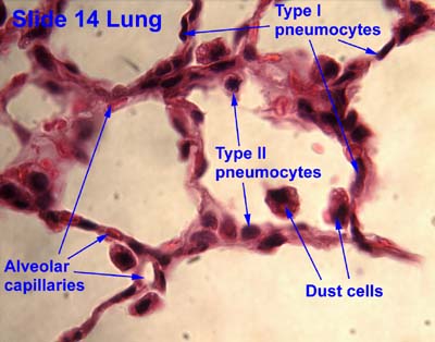
Figure 5: Electron Micrograph of Alveolar Cells (Retrieved from
No comments:
Post a Comment
Note: Only a member of this blog may post a comment.